Abstract
Clinical Image
58-year-old male patient who came to the dermatology service for a clinical picture
Francisco Javier Torres-Gómez*
Published: 30 April, 2021 | Volume 5 - Issue 1 | Pages: 018-019
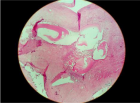
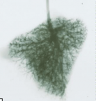
58-year-old male patient who came to the dermatology service for a clinical picture consisting of generalized erythematous scaly and pruritic lesions of 2 years of evolution. The clinical judgments provided were: pityriasis versicolor, drop psoriasis, pityriasis rubra pilaris and secondary syphilis (without serology confirming this last hypothesis then). A biopsy of a lesion located on the right costal side was performed. The serology was negative in a second time.
Read Full Article HTML DOI: 10.29328/journal.adr.1001016 Cite this Article Read Full Article PDF
References
- Varada S, Dabade T, Loo DS. Uncommon presentations of tinea versicolor. Dermatol Pract Concept. 2014; 4: 93-96. PubMed: https://pubmed.ncbi.nlm.nih.gov/25126470/
- Ferry M, Shedlofsky L, Newman A, Mengesha Y, Blumetti B. Tinea InVersicolor: A Rare Distribution of a Common Eruption. Cureus. 2020; 12: e6689. PubMed: https://pubmed.ncbi.nlm.nih.gov/32104626/
Figures:
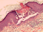
Figure 1
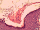
Figure 2
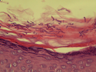
Figure 3
Similar Articles
-
Clinical significance of Vibration Anesthesia on reducing pain of Ring-Block (Subcutaneous Injections) in the patients undergoing Hair Restoration SurgeryMuhammad Ahmad*,Mohammad Humayun Mohmand. Clinical significance of Vibration Anesthesia on reducing pain of Ring-Block (Subcutaneous Injections) in the patients undergoing Hair Restoration Surgery. . 2017 doi: 10.29328/journal.adr.1001001; 1: 001-005
-
Lifestyle Diseases and the Hair Growth Cycle: A multidisciplinary approach using Nourkrin® with Marilex®, a proteoglycan replacement therapy, for anagen induction and maintenanceThom E*,Thom EW. Lifestyle Diseases and the Hair Growth Cycle: A multidisciplinary approach using Nourkrin® with Marilex®, a proteoglycan replacement therapy, for anagen induction and maintenance. . 2017 doi: 10.29328/journal.adr.1001002; 1: 006-011
-
It is not invisible! A case report of 2 patients with scalp Lichen Planopilaris mimicking Androgenic AlopeciaFabio Rinaldi*,Sorbellini Elisabetta,Pinto Daniela,Marzani Barbara. It is not invisible! A case report of 2 patients with scalp Lichen Planopilaris mimicking Androgenic Alopecia. . 2017 doi: 10.29328/journal.adr.1001003; 1: 012-017
-
Metabolic Syndrome, Cardiovascular Disease and the Hair Growth Cycle: Addressing hair growth disruptions using Nourkrin® with Marilex® as a proteoglycan replacement therapy: A concise reviewThom E*,Wadstein J,Kingsley DH*,Thom EW . Metabolic Syndrome, Cardiovascular Disease and the Hair Growth Cycle: Addressing hair growth disruptions using Nourkrin® with Marilex® as a proteoglycan replacement therapy: A concise review. . 2018 doi: 10.29328/journal.adr.1001004; 2: 001-007
-
Linear IgA bullous dermatosis in a child successfully responding to oral antibioticsG Senhaji*,H Bay Bay,O El Jouari,A Lamouaffaq,Z Douhi,S Elloudi,FZ Mernissi ,R Dassouli. Linear IgA bullous dermatosis in a child successfully responding to oral antibiotics . . 2018 doi: 10.29328/journal.adr.1001005; 2: 008-011
-
Daub, Discolouration, Pigmentation-Solar LentigoAnubha Bajaj*. Daub, Discolouration, Pigmentation-Solar Lentigo. . 2019 doi: 10.29328/journal.adr.1001006; 3: 001-006
-
We may need to reconsider when to apply sunscreen in our daily lifeWin L Chiou*. We may need to reconsider when to apply sunscreen in our daily life. . 2019 doi: 10.29328/journal.adr.1001007; 3: 007-010
-
Evaluation in real life of the impact of photo-protection counseling in patients with actinic keratosisCharles Taïeb*,Khaled Ezzedine,Sophie Seité. Evaluation in real life of the impact of photo-protection counseling in patients with actinic keratosis. . 2019 doi: 10.29328/journal.adr.1001008; 3: 011-012
-
Leprosy persistence in the health district of Kenieba despite its elimination as a public health problem at the national level in MaliIlo Dicko*,Yaya Ibrahim Coulibaly,Modibo Keita,Housseini Dolo,Modibo Sangaré,Abdoulaye Fomba,Mamoudou Kodio,Nouhou Diarra,Mamadou Sidibé,Oumar Maiga,Moussa Brema Sangaré,Siaka Yamoussa Coulibaly,Michel Emmanuel Coulibaly,Mamadou Dolo,Sory Ibrahima Fomba,Abdallah Amadou Diallo,Floribert Fossuo Thotchum,Ousmane Faye,Samba Ousmane Sow. Leprosy persistence in the health district of Kenieba despite its elimination as a public health problem at the national level in Mali . . 2020 doi: 10.29328/journal.adr.1001009; 4: 001-005
-
Bee venom: a case of effectiveness on skin varicosities veins with review of its dermatological benefitsMohamed El Amraoui*,Rachid Frikh,Naoufal Hjira,Mohammed Boui. Bee venom: a case of effectiveness on skin varicosities veins with review of its dermatological benefits. . 2020 doi: 10.29328/journal.adr.1001010; 4: 006-008
Recently Viewed
-
Obesity in Patients with Chronic Obstructive Pulmonary Disease as a Separate Clinical PhenotypeDaria A Prokonich*, Tatiana V Saprina, Ekaterina B Bukreeva. Obesity in Patients with Chronic Obstructive Pulmonary Disease as a Separate Clinical Phenotype. J Pulmonol Respir Res. 2024: doi: 10.29328/journal.jprr.1001060; 8: 053-055
-
Current Practices for Severe Alpha-1 Antitrypsin Deficiency Associated COPD and EmphysemaMJ Nicholson*, M Seigo. Current Practices for Severe Alpha-1 Antitrypsin Deficiency Associated COPD and Emphysema. J Pulmonol Respir Res. 2024: doi: 10.29328/journal.jprr.1001058; 8: 044-047
-
Navigating Neurodegenerative Disorders: A Comprehensive Review of Current and Emerging Therapies for Neurodegenerative DisordersShashikant Kharat*, Sanjana Mali*, Gayatri Korade, Rakhi Gaykar. Navigating Neurodegenerative Disorders: A Comprehensive Review of Current and Emerging Therapies for Neurodegenerative Disorders. J Neurosci Neurol Disord. 2024: doi: 10.29328/journal.jnnd.1001095; 8: 033-046
-
Metastatic Brain Melanoma: A Rare Case with Review of LiteratureNeha Singh,Gaurav Raj,Akshay Kumar,Deepak Kumar Singh,Shivansh Dixit,Kaustubh Gupta*. Metastatic Brain Melanoma: A Rare Case with Review of Literature. J Radiol Oncol. 2025: doi: 10.29328/journal.jro.1001080; 9: 050-053
-
Validation of Prognostic Scores for Attempted Vaginal Delivery in Scar UterusMouiman Soukaina*,Mourran Oumaima,Etber Amina,Zeraidi Najia,Slaoui Aziz,Baydada Aziz. Validation of Prognostic Scores for Attempted Vaginal Delivery in Scar Uterus. Clin J Obstet Gynecol. 2025: doi: 10.29328/journal.cjog.1001185; 8: 023-029
Most Viewed
-
Evaluation of Biostimulants Based on Recovered Protein Hydrolysates from Animal By-products as Plant Growth EnhancersH Pérez-Aguilar*, M Lacruz-Asaro, F Arán-Ais. Evaluation of Biostimulants Based on Recovered Protein Hydrolysates from Animal By-products as Plant Growth Enhancers. J Plant Sci Phytopathol. 2023 doi: 10.29328/journal.jpsp.1001104; 7: 042-047
-
Sinonasal Myxoma Extending into the Orbit in a 4-Year Old: A Case PresentationJulian A Purrinos*, Ramzi Younis. Sinonasal Myxoma Extending into the Orbit in a 4-Year Old: A Case Presentation. Arch Case Rep. 2024 doi: 10.29328/journal.acr.1001099; 8: 075-077
-
Feasibility study of magnetic sensing for detecting single-neuron action potentialsDenis Tonini,Kai Wu,Renata Saha,Jian-Ping Wang*. Feasibility study of magnetic sensing for detecting single-neuron action potentials. Ann Biomed Sci Eng. 2022 doi: 10.29328/journal.abse.1001018; 6: 019-029
-
Pediatric Dysgerminoma: Unveiling a Rare Ovarian TumorFaten Limaiem*, Khalil Saffar, Ahmed Halouani. Pediatric Dysgerminoma: Unveiling a Rare Ovarian Tumor. Arch Case Rep. 2024 doi: 10.29328/journal.acr.1001087; 8: 010-013
-
Physical activity can change the physiological and psychological circumstances during COVID-19 pandemic: A narrative reviewKhashayar Maroufi*. Physical activity can change the physiological and psychological circumstances during COVID-19 pandemic: A narrative review. J Sports Med Ther. 2021 doi: 10.29328/journal.jsmt.1001051; 6: 001-007

HSPI: We're glad you're here. Please click "create a new Query" if you are a new visitor to our website and need further information from us.
If you are already a member of our network and need to keep track of any developments regarding a question you have already submitted, click "take me to my Query."


