Abstract
Research Article
Artemisia Naphta: A novel oil extract for sensitive and acne prone skin
Edwardo Perez*, Kan Tao, Lili Guo, Jose Fernandez, Corey Webb, Junfeng Liu, Xincheng Hu and Dan Yang
Published: 15 June, 2021 | Volume 5 - Issue 1 | Pages: 022-029
Background: The plant Artemisia annua has been used in traditional Chinese medicine for many years. Rich in bioactive molecules, the A. annua plant is used to extract the anti-malaria compound artemisinin (< 1%), which results in most of the plant being unutilized. One byproduct of artemisinin extraction is artemisia naphtha (AN), which has yet to be studied extensively.
Aims: Study the activity of a novel AN oil extract against microbes, pro-inflammatory cytokines, and dermatological endpoints that are key for eczema and acne pathogenesis to determine if an effective A. annua extract for these skin conditions can be developed.
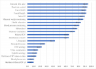
Methods: Gas chromatography-mass spectrometry was performed to determine the composition of AN oil. P. acnes, S. aureus, M. furfur, and C. albicans were cultured to determine minimal inhibitory concentration. in vitro studies utilizing keratinocytes and macrophages were treated with AN oil and gene expression measured by quantitative RT-PCR. A 13-subject clinical trial was performed with 1% AN oil Gel to assess its potential benefits for sensitive and acne prone skin.
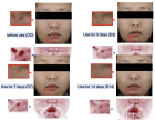
Results: AN oil upregulates filaggrin gene expression and possesses antimicrobial and anti-inflammatory activity inhibiting LPS, S. aureus and "Th2 induced" pro-inflammatory mediator release (IL-6, IL-8 and TSLP). Clinical assessment of 1% AN Gel shows it reduces acne blemishes and the appearance of redness.
Conclusion: Previously an underutilized and unpurified byproduct, AN is now the source to develop the first topical AN oil for cosmetic use with an activity profile that suggests it is effective for those with sensitive and/or acne prone skin.
Read Full Article HTML DOI: 10.29328/journal.adr.1001018 Cite this Article Read Full Article PDF
Keywords:
Anti-inflammatory; Antimicrobial; Acne; Eczema; Sensitive skin; Cosmetic
References
- Mueller MS, Karhagomba IB, Hirt HM, Wemakor E. The potential of Artemisia annua L. as a locally produced remedy for malaria in the tropics: agricultural, chemical and clinical aspects. J Ethnopharmacol. 2000; 73: 487-493. https://pubmed.ncbi.nlm.nih.gov/11091003/
- Septembre-Malaterre A, Rakoto ML, Marodon C, Bedoui Y, Jessica Nakab et al. Artemisia annua, a Traditional Plant Brought to Light. Int J Mol Sci. 2020; 21: 4986. https://pubmed.ncbi.nlm.nih.gov/32679734/
- Li T, Chen H, Wei N, Mei X, Zhang S, et al. Anti-inflammatory and immunomodulatory mechanisms of artemisinin on contact hypersensitivity. Int Immunopharmacol. 2012; 12: 144-150. https://pubmed.ncbi.nlm.nih.gov/22122827/
- Xue X, Dong Z, Deng Y, Yin S, Wang P, et al. [Dihydroartemisinin alleviates atopic dermatitis in mice by inhibiting mast cell infiltration]. Nan Fang Yi Ke Da Xue Xue Bao. 2020; 40: 1480-1487. https://pubmed.ncbi.nlm.nih.gov/33118501/
- Li YJ, Guo Y, Yang Q Weng XG, Yang L, et al. Flavonoids casticin and chrysosplenol D from Artemisia annua L. inhibit inflammation in vitro and in vivo. Toxicol Appl Pharmacol. 2015; 286: 151-158. https://pubmed.ncbi.nlm.nih.gov/25891417/
- Ciftci ON, Cahyadi J, Guigard SE, Saldaña MDA. Optimization of artemisinin extraction from Artemisia annua L. with supercritical carbon dioxide + ethanol using response surface methodology. Electrophoresis. 2018. https://pubmed.ncbi.nlm.nih.gov/29756212/
- Yu J, Wang G, Jiang N. Study on the repairing effect of cosmetics containing Artemisia annua on sensitive skin. J Cosmetics Dermatol Sci Appli. 2020; 10: 8-19.
- Wu C, Yan Y, Wang Y, Sun P, Qi R. Antibacterial epoxy composites with addition of natural Artemisisa annua waste. e-Polymers. 2020; 20: 262-271.
- Saify ZS, Ahsan O, Dayo A. Cineole as skin penetration enhancer. Pak J Pharm Sci. 2000; 13: 29-32. https://pubmed.ncbi.nlm.nih.gov/16414836/
- Tran TA, Ho MT, Song YW, Cho M, Cho SK. Camphor Induces Proliferative and Anti-senescence Activities in Human Primary Dermal Fibroblasts and Inhibits UV-Induced Wrinkle Formation in Mouse Skin. Phytother Res. 2015; 29: 1917-1925. https://pubmed.ncbi.nlm.nih.gov/26458283/
- Quintans-Junior L, Moreira JCF, Pasquali MAB, Rabie SMS, Pires AS, et al. Antinociceptive Activity and Redox Profile of the Monoterpenes (+)-Camphene, p-Cymene, and Geranyl Acetate in Experimental Models. ISRN Toxicol. 2013; 2013: 459530. https://pubmed.ncbi.nlm.nih.gov/23724298/
- Marchese A, Arciola CR, Barbieri R, Silva AS, Nabavi SF, et al. Update on Monoterpenes as Antimicrobial Agents: A Particular Focus on p-Cymene. Materials (Basel). 2017; 10: 947. https://pubmed.ncbi.nlm.nih.gov/28809799/
- Luna A, Tarifa MF, Fernandez ME, Caliva JE, Pellegrini S, et al. Thymol, alpha tocopherol, and ascorbyl palmitate supplementation as growth enhancers for broiler chickens. Poult Sci. 2019; 98: 1012-1016. https://pubmed.ncbi.nlm.nih.gov/30165460/
- Scholz CFP, Kilian M. The natural history of cutaneous propionibacteria, and reclassification of selected species within the genus Propionibacterium to the proposed novel genera Acidipropionibacterium gen. nov, Cutibacterium gen. nov. and Pseudopropionibacterium gen. nov. Int J Syst Evol Microbiol. 2016; 66: 4422-4432. https://pubmed.ncbi.nlm.nih.gov/27488827/
- Hauser C, Wuethrich B, Matter L, Wilhelm JA, Schopfer K. The immune response to S. aureus in atopic dermatitis. Acta Derm Venereol Suppl (Stockh). 1985; 114: 101-104. https://pubmed.ncbi.nlm.nih.gov/3859157/
- Liu CH, Zou WX, Lu H, Tan RX. Antifungal activity of Artemisia annua endophyte cultures against phytopathogenic fungi. J Biotechnol. 2001; 88: 277-282. https://pubmed.ncbi.nlm.nih.gov/11434973/
- Jugeau S, Tenaud I, Knol AC, Jarrousse V, Quereux G, et al. Induction of toll-like receptors by Propionibacterium acnes. Br J Dermatol. 2005; 153: 1105-1113. https://pubmed.ncbi.nlm.nih.gov/16307644/
- Gong JQ, Lin L, Lin T, Hao F, Zeng FQ, et al. Skin colonization by Staphylococcus aureus in patients with eczema and atopic dermatitis and relevant combined topical therapy: a double-blind multicentre randomized controlled trial. Br J Dermatol. 2006; 155: 680-687. https://pubmed.ncbi.nlm.nih.gov/16965415/
- Blicharz L, Usarek P, Młynarczyk G, Skowroński K, Rudnicka L, et al. Is Itch Intensity in Atopic Dermatitis Associated with Skin Colonization by Staphylococcus aureus? Indian J Dermatol. 2020; 65: 17-21. https://pubmed.ncbi.nlm.nih.gov/32029934/
- Turner MJ, Zhou B. A new itch to scratch for TSLP. Trends Immunol. 2014; 35: 49-50. https://pubmed.ncbi.nlm.nih.gov/24412411/
- Leung DY, Boguniewicz M, Howell MD, Nomura I, Hamid QA. New insights into atopic dermatitis. J Clin Invest, 2004. 113: 651-657. https://pubmed.ncbi.nlm.nih.gov/14991059/
- Levin J, Friedlander SF, Del Rosso JQ. Atopic dermatitis and the stratum corneum: part 1: the role of filaggrin in the stratum corneum barrier and atopic skin. J Clin Aesthet Dermatol. 2013; 6: 16-22. https://pubmed.ncbi.nlm.nih.gov/24155988/
Figures:

Figure 1

Figure 2

Figure 3

Figure 4

Figure 5
Similar Articles
-
Clinical significance of Vibration Anesthesia on reducing pain of Ring-Block (Subcutaneous Injections) in the patients undergoing Hair Restoration SurgeryMuhammad Ahmad*,Mohammad Humayun Mohmand. Clinical significance of Vibration Anesthesia on reducing pain of Ring-Block (Subcutaneous Injections) in the patients undergoing Hair Restoration Surgery. . 2017 doi: 10.29328/journal.adr.1001001; 1: 001-005
-
Artemisia Naphta: A novel oil extract for sensitive and acne prone skinEdwardo Perez*,Kan Tao,Lili Guo,Jose Fernandez,Corey Webb,Junfeng Liu,Xincheng Hu,Dan Yang. Artemisia Naphta: A novel oil extract for sensitive and acne prone skin. . 2021 doi: 10.29328/journal.adr.1001018; 5: 022-029
Recently Viewed
-
Investigation of Stain Patterns from Diverse Blood Samples on Various SurfacesSonia Rajkumari*. Investigation of Stain Patterns from Diverse Blood Samples on Various Surfaces. J Forensic Sci Res. 2024: doi: 10.29328/journal.jfsr.1001061; 8: 028034
-
The Impact of Forensic Science on the Legal System in IndiaSrishti*. The Impact of Forensic Science on the Legal System in India. J Forensic Sci Res. 2025: doi: 10.29328/journal.jfsr.1001072; 9: 001-006
-
Postural Stability Induced by Supervised Physical Training may improve also Oxygen Cost of Exercise and Walking Capacity in Post-Menopause, Obese WomenFernanda Velluzzi,Massimiliano Pau,Andrea Loviselli,Raffaele Milia,Daniela Lai,Daniele Concu,Gianmarco Angius,Abdallah Raweh,Andrea Fois,Alberto Concu*. Postural Stability Induced by Supervised Physical Training may improve also Oxygen Cost of Exercise and Walking Capacity in Post-Menopause, Obese Women. J Nov Physiother Rehabil. 2017: doi: 10.29328/journal.jnpr.1001001; 1: 001-011
-
Antibacterial Screening of Lippia origanoides Essential Oil on Gram-negative BacteriaRodrigo Marcelino Zacarias de Andrade, Bernardina de Paixão Santos, Roberson Matteus Fernandes Silva, Mateus Gonçalves Silva*, Igor de Sousa Oliveira, Sávio Benvindo Ferreira, Rafaelle Cavalcante Lira. Antibacterial Screening of Lippia origanoides Essential Oil on Gram-negative Bacteria. Arch Pharm Pharma Sci. 2024: doi: 10.29328/journal.apps.1001053; 8: 024-028.
-
Recovery of craniofacial proportions using the Nuvola Op System protocolAlessandro Carrafiello*. Recovery of craniofacial proportions using the Nuvola Op System protocol. J Oral Health Craniofac Sci. 2022: doi: 10.29328/journal.johcs.1001041; 7: 022-026
Most Viewed
-
Evaluation of Biostimulants Based on Recovered Protein Hydrolysates from Animal By-products as Plant Growth EnhancersH Pérez-Aguilar*, M Lacruz-Asaro, F Arán-Ais. Evaluation of Biostimulants Based on Recovered Protein Hydrolysates from Animal By-products as Plant Growth Enhancers. J Plant Sci Phytopathol. 2023 doi: 10.29328/journal.jpsp.1001104; 7: 042-047
-
Sinonasal Myxoma Extending into the Orbit in a 4-Year Old: A Case PresentationJulian A Purrinos*, Ramzi Younis. Sinonasal Myxoma Extending into the Orbit in a 4-Year Old: A Case Presentation. Arch Case Rep. 2024 doi: 10.29328/journal.acr.1001099; 8: 075-077
-
Feasibility study of magnetic sensing for detecting single-neuron action potentialsDenis Tonini,Kai Wu,Renata Saha,Jian-Ping Wang*. Feasibility study of magnetic sensing for detecting single-neuron action potentials. Ann Biomed Sci Eng. 2022 doi: 10.29328/journal.abse.1001018; 6: 019-029
-
Pediatric Dysgerminoma: Unveiling a Rare Ovarian TumorFaten Limaiem*, Khalil Saffar, Ahmed Halouani. Pediatric Dysgerminoma: Unveiling a Rare Ovarian Tumor. Arch Case Rep. 2024 doi: 10.29328/journal.acr.1001087; 8: 010-013
-
Physical activity can change the physiological and psychological circumstances during COVID-19 pandemic: A narrative reviewKhashayar Maroufi*. Physical activity can change the physiological and psychological circumstances during COVID-19 pandemic: A narrative review. J Sports Med Ther. 2021 doi: 10.29328/journal.jsmt.1001051; 6: 001-007

HSPI: We're glad you're here. Please click "create a new Query" if you are a new visitor to our website and need further information from us.
If you are already a member of our network and need to keep track of any developments regarding a question you have already submitted, click "take me to my Query."


